- This event has passed.
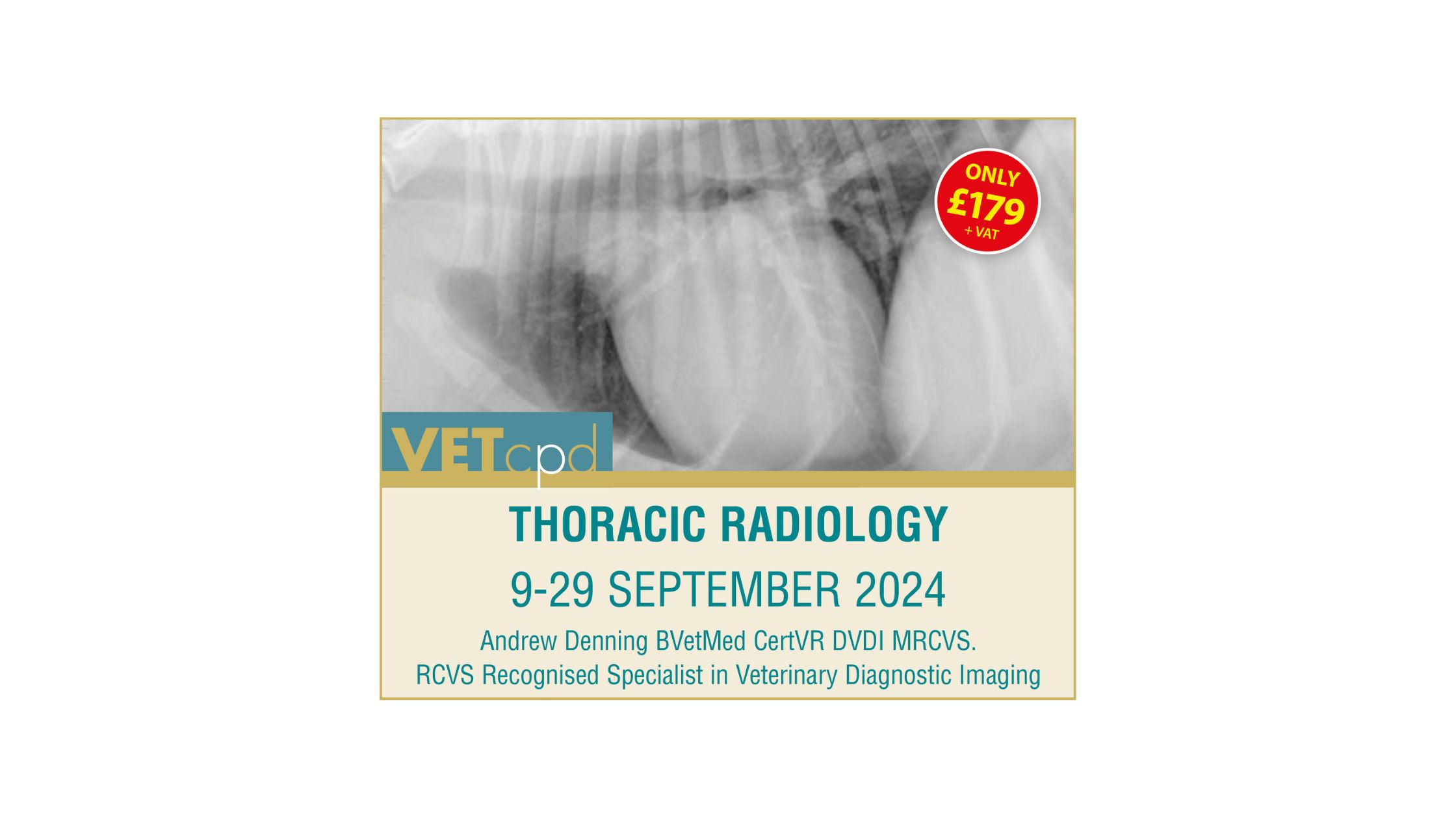
Thoracic Radiology in Dogs and Cats
9th September 2024 - 29th September 2024
£179
Course Overview
Thoracic imaging is one of the most common diagnostic techniques performed in clinical practice. In most clinical settings, radiography remains the main imaging modality for the thorax. Ultrasound can also provide useful information. CT is becoming more widely available.
Fluroscopy will not be considered in this course but is useful for swallowing studies.
For any imaging study it is necessary to identify the clinical questions that need to be answered and then to specify the area to be imaged and the appropriate technique to be used. For example investigation of the thorax for symptoms of cough or dyspnoea or screening of the thorax for thoracic metastasis will require three-view radiographs of the thorax or thoracic CT. It is not possible to cover the whole subject of thoracic radiology in a course of this type and length but I hope to go through the basic principles so that you can then apply these to your imaging practice in the future.


Leave a Reply